Tablet enters operating theatre
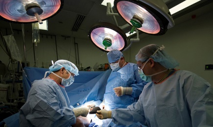
On August 15, the surgical team at the Asklepios Klinik Barmbek in Hamburg successfully tested the new Fraunhofer-app during a liver operation. A liver cancer operation usually lasts many hours because the organ is difficult to operate. It hosts a branching vessel structure through which one and a half liters of blood flow every minute. If a surgeon makes a cut in an inappropriate place, this puts the patient at risk of severe blood loss.
In addition, doctors must ensure that the patient retains enough organ volume for survival and that this volume is sufficiently supplied with blood. To accomplish this, doctors need to know as accurately as possible both before and during an operation where blood vessels inside the organ are located.
Where are those blood vessels?
The new tablet app from the Fraunhofer Institute for Medical Image Computing MEVIS in Bremen promises to deliver this support. It is based on the established MEVIS software for liver operation planning that is employed in clinics worldwide and has been used for more than 6000 patients. Based on 3D x-ray images, the software can reconstruct the locations of blood vessels in the liver for each patient. Before an operation, surgeons can then precisely plan how and where to place the scalpel to most effectively remove a tumor.
However, there are limitations: doctors usually have little opportunity to view the software images during surgery and compare the surgical situation with planning data. Some surgeons even print out images to take into the operating room. “With our app, the entire set of planning data can be shown directly on the operating table” said MEVIS computer scientist Alexander Köhn.
Virtually looking into the organ
At the intervention in Hamburg, the clinicians used a further feature of the new app. With the integrated camera, the tablet could film the liver during the operation. The app then superimposed the planning data – a branched network showing the vessel system in different colors. “Using this function, we can virtually look into the organ and make the tumor and vessel structures visible” said Prof. Dr. Karl Oldhafer, Chief of the Department of Surgery at the Asklepios Klinik Barmbek in Hamburg.
This simplifies comparison to determine whether the intervention has gone according to plan. “With this new technology, we are able to better implement computer-supported operation planning for tumor removal” remarked Oldhafer. “The method has great potential. We imagine using it for operations on other organs, such as the pancreas.”
Clever app
For future interventions, the app offers more capabilities. By simply marking the touchscreen, doctors can measure the length of a vessel to be removed. This helps the doctor estimate whether the remaining ends can be sewn together or whether a new piece of vessel must be inserted.
After the surgeon removes certain vessels, he can remove them on the app screen with a virtual ‘eraser’. The separated vessels disappear from the screen and let the doctor view underlying structures. If, during the operation, a tumor is judged to be larger than at first thought, surgeons must make snap decisions. The MEVIS app can also help here. If additional vessels must be removed, the app calculates which parts of the liver will no longer be sufficiently supplied with blood. This lets the surgeon better estimate whether the remaining organ volume is large enough for the patient to survive.